


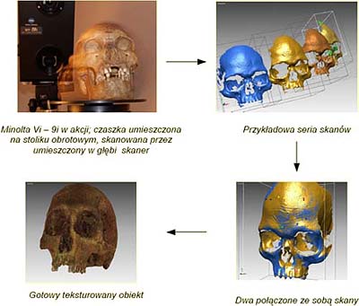
3D Scanner
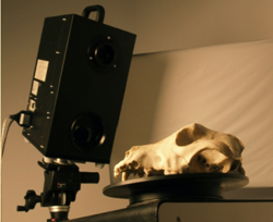 |
Minolta VI - 9i 3D Laser Scanner This apparatus is capable of transfer ring a three-dimensional image of a given object (of defined parameters – see limitations) to computer memory. The scanner operates on the principle of laser triangulation in which the object is scanned with the aid of a line of laser light whose course is followed by a digital camera. The curvature of the line on the object under study is used as the basis for building up the three-dimensional image. The camera also reads off the colour of the object, which can be added to the model in the form of texture. The object for scanning is placed on a special table and read with the laser as the apparatus rotates through a determined number of degrees. The individual scans are then combined together by reference to characteristic points determined by the operator. |
Possibilities
The scanner does not harm the eyes and so may be used to scan human faces. Depending on the size of the object, it is possible to use one of three objectives whose accuracies are as follows:
TELE XYZ: ±0.05mm/ ±0.10mm
MIDDLE XYZ ±0.10mm/ ±0.20mm
WIDE XYZ ±0.20mm/ ±0.40mm
Properties of scanned objects - limitations
- Transparent surfaces or those reflecting laser light must be covered with powder
- Deep-black surfaces are the ideal background colour for the scanner and must also be made subject to defined processes.
- Objects of dimensions greater than 1200/900/750 mm have to be scanned in stages.
- Objects of very complex shapes and sizes over 2 cm must be considered separately.
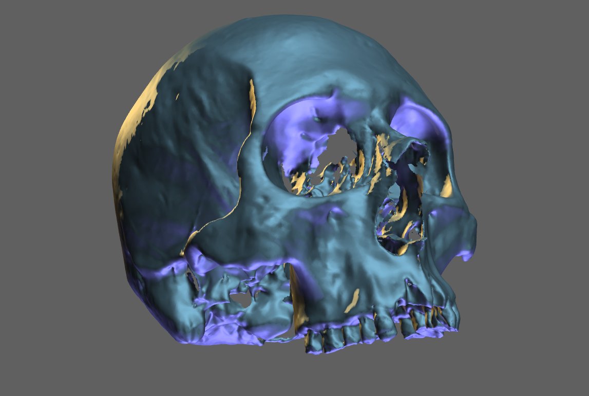
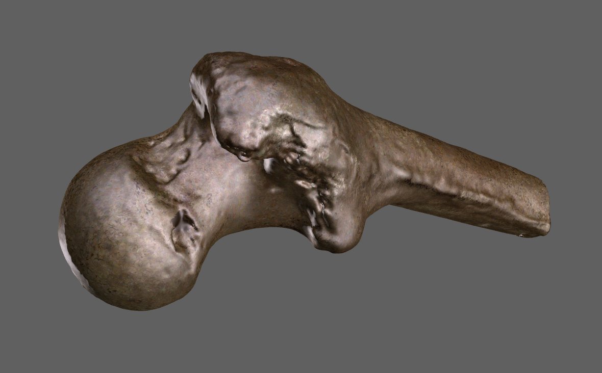
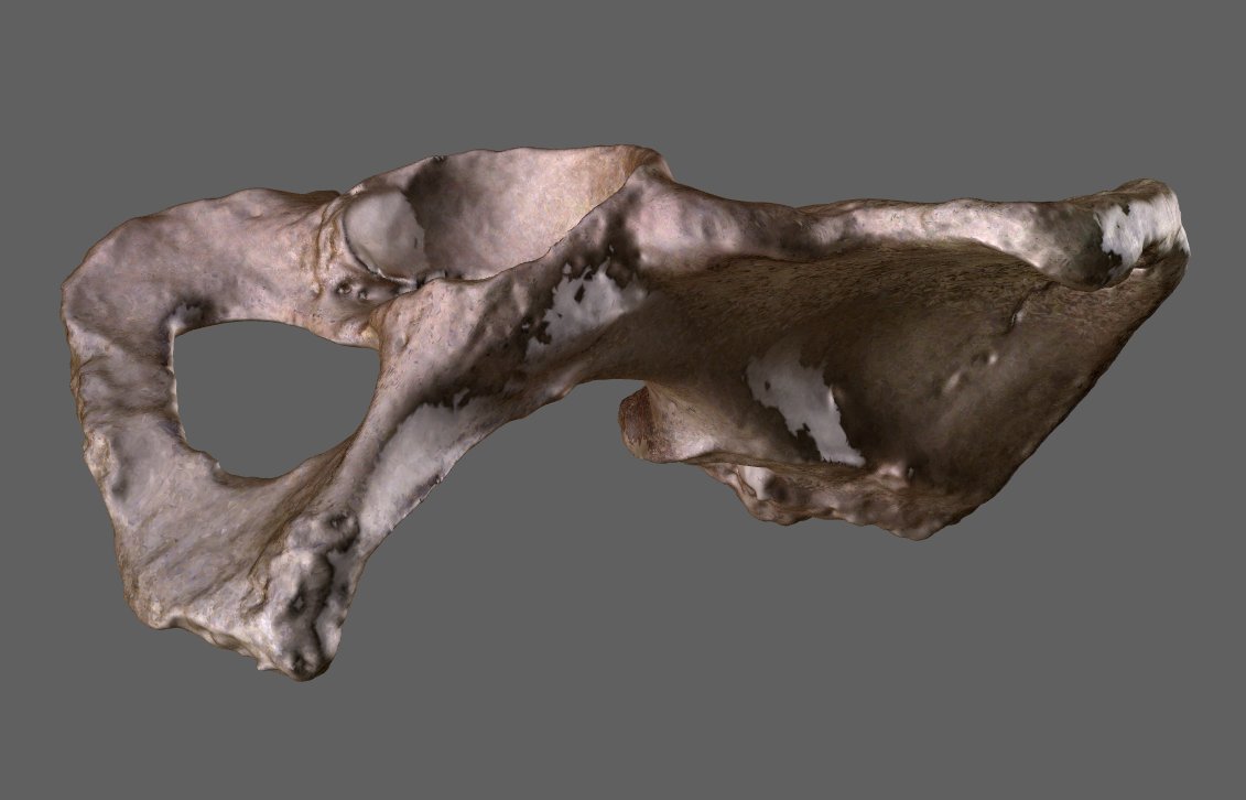
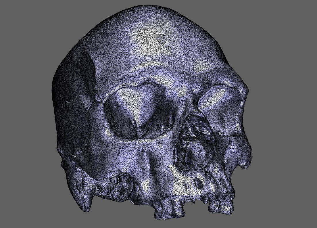
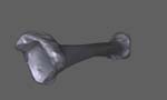
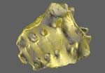
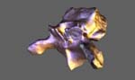
Sample scans:
 |
 |
 |
 |
 |
 |
 |
 |
 |
3D - PDF files to download
 |
 |
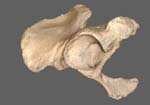 |
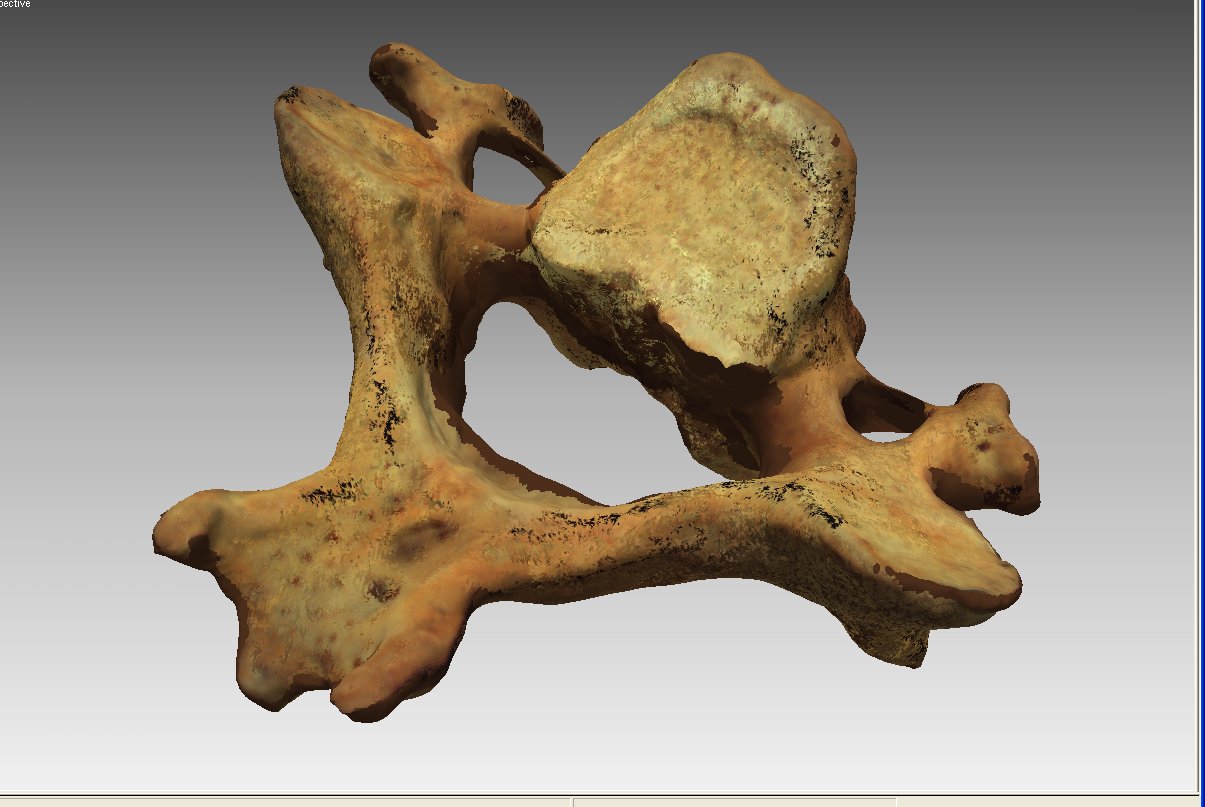
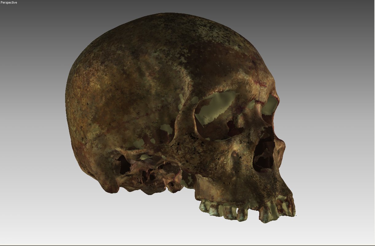
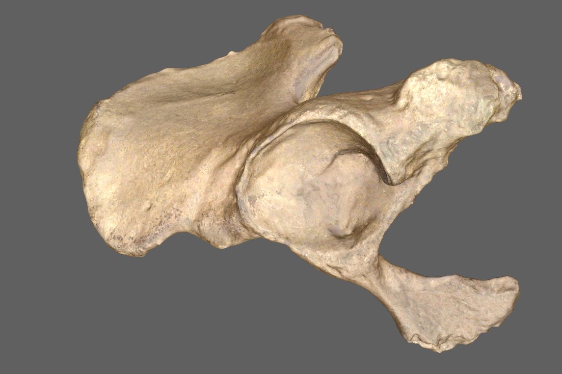
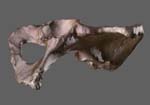
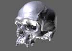
| Pelvis bone (10,0 MB) | Skull (6,5 MB) | Pelvis bone (9,2 MB) |
 |
 |
 |
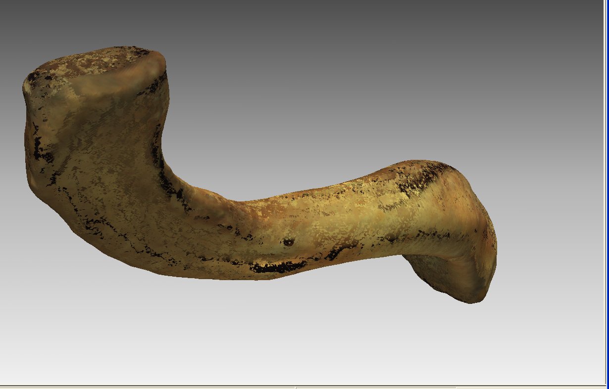
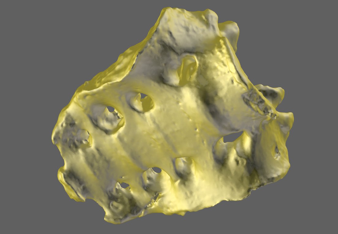
| Tiba (3,7 MB) | Sacral bone (4,3 MB) | Vertebra (2,3 MB) |
HITACHI S-3400N Scanning Electron Microscope
|
|
HITACHI S-3400N Scanning Electron Microscope Operating in variable-vacuum conditions and equipped with an EDS x-ray microanalyser, this apparatus provides for the observation and both quantitative and qualitative analysis of metal preparations and non-metal ones that conduct electricity, as well as biological samples, and non-conducting materials Main parameters of the microscope:
The microscope is also equipped with:
|


Research work is carried out for both the Museum and Institute of Zoology and other scientific institutes, higher education establishments, industrial partners, and so on.
Those interested in the research opportunities are invited to contact:
tel. 022 357-50-01
022 357-30-01
e-mail: This email address is being protected from spambots. You need JavaScript enabled to view it.
Examples:

 |
 |
 |
| 1DC k_m01 | Enicocephalidae | F pressilabris samiec |
 |
 |
 |
| Kokolit 1 | Kokolit 2 | Trissonchulus benepapillosus |
 |
 |
 |
| Oko komara | Warstwa fosoranowa na AL | Trissonchulus benepapillosus |
 |
 |
 |
| Fragment przewodu pokarmowego ślimaka Ceapea vindobonensis | Fragment przewodu pokarmowego ślimaka Ceapea vindobonensis | (obraz SE) Ochterus |
 |
 |
 |
| Fragment skrzydła Borysthenes | Patyczak | Warstwa Ni |
 |
 |
| Opoka - okolice Bychawy | Skorupa ślimaka - Helicidae: Cepaea |
 |
 |
| Skorupa ślimaka - Helicidae: Cepaea |
Warstwa Ni na warstwie NI-P (podłoże Cu) |